Account
Inquiry
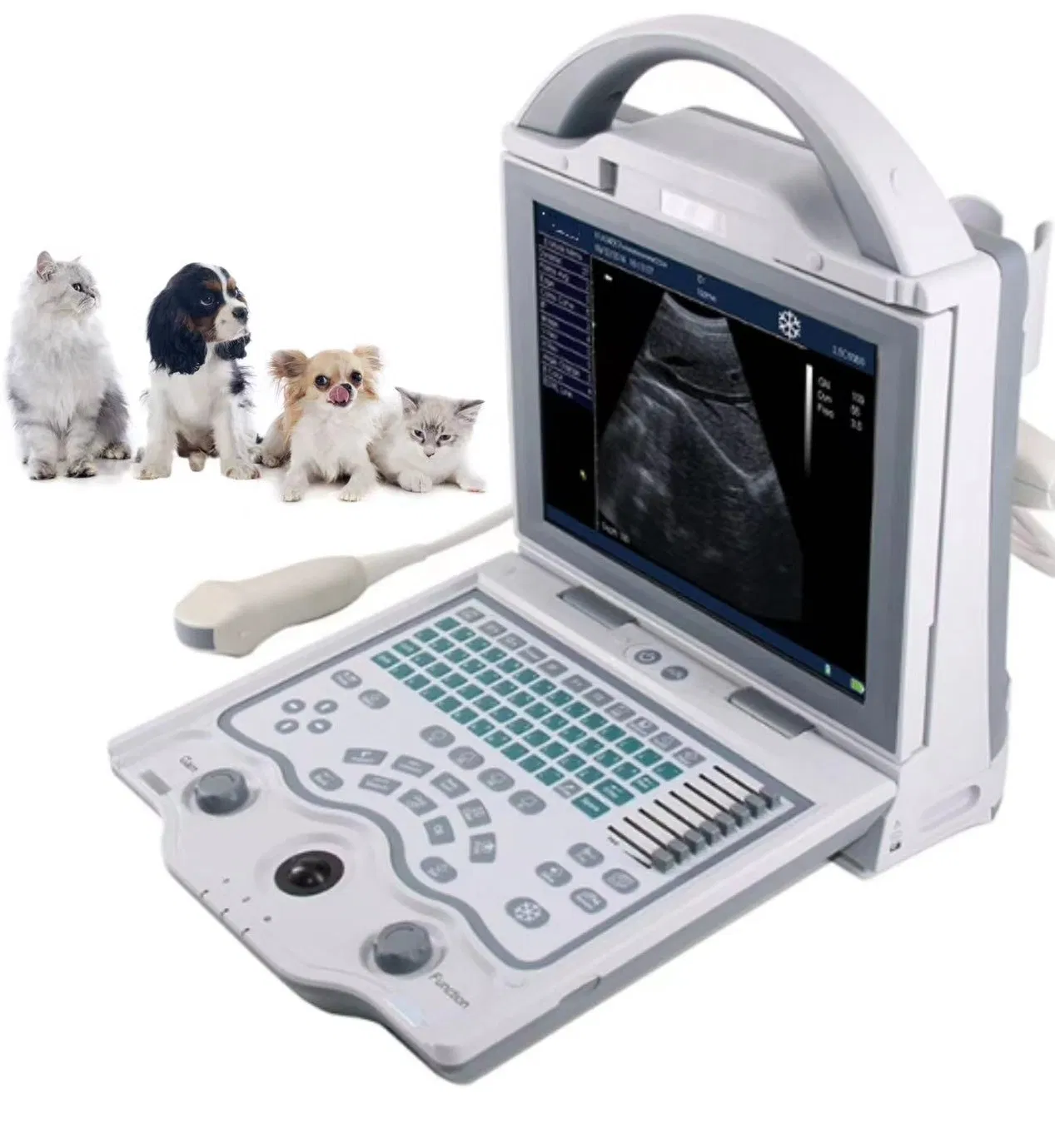
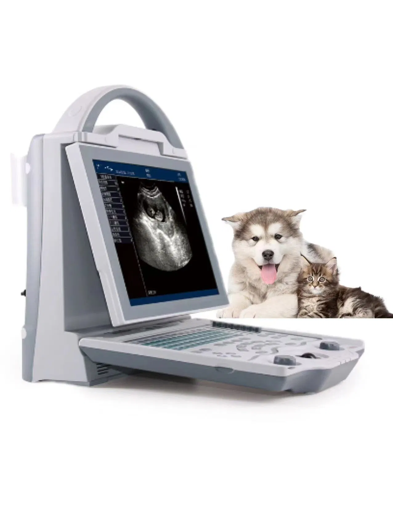
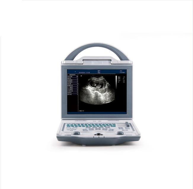
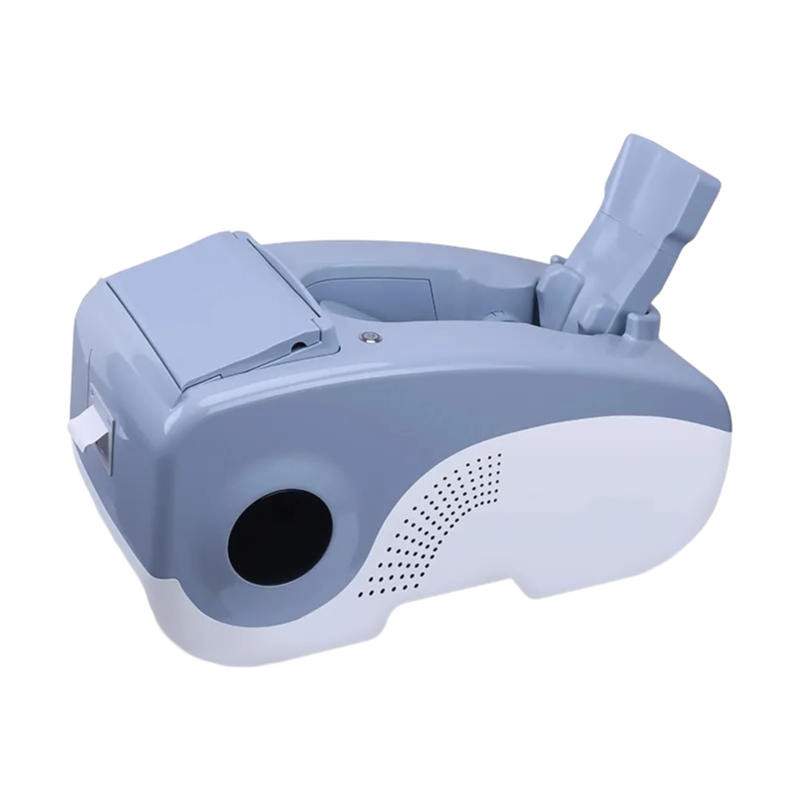
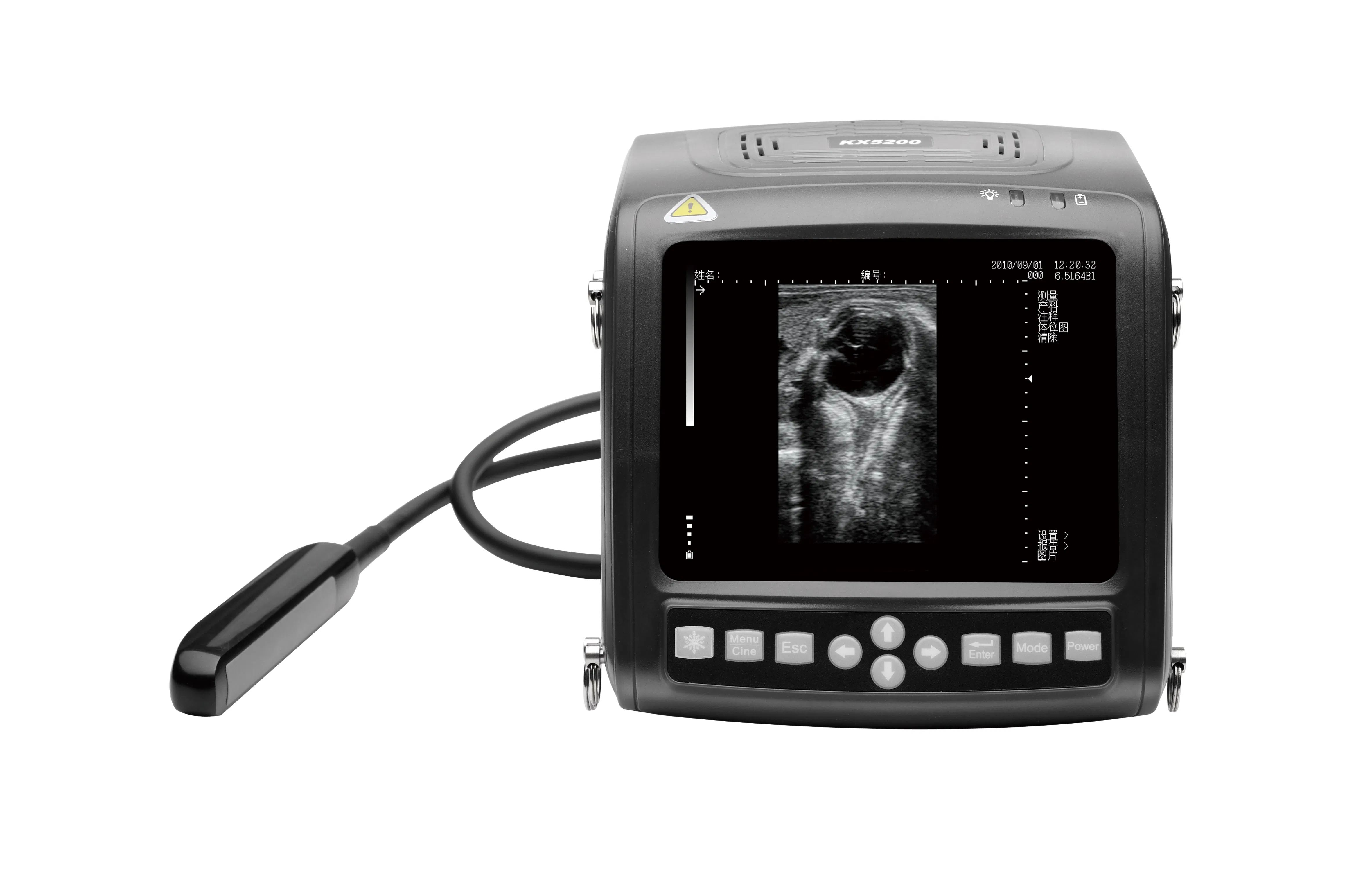
Scrolling through the saved OB dataset from last week's late-term bitch scan, it's pretty clear the uterine wall echoes are falling off at 180mm depth, you'll need that 8-step TGC to tweak the gain gradient—most buyers in mixed practice go straight to the 6.5MHz micro-convex for deep abdominal work.
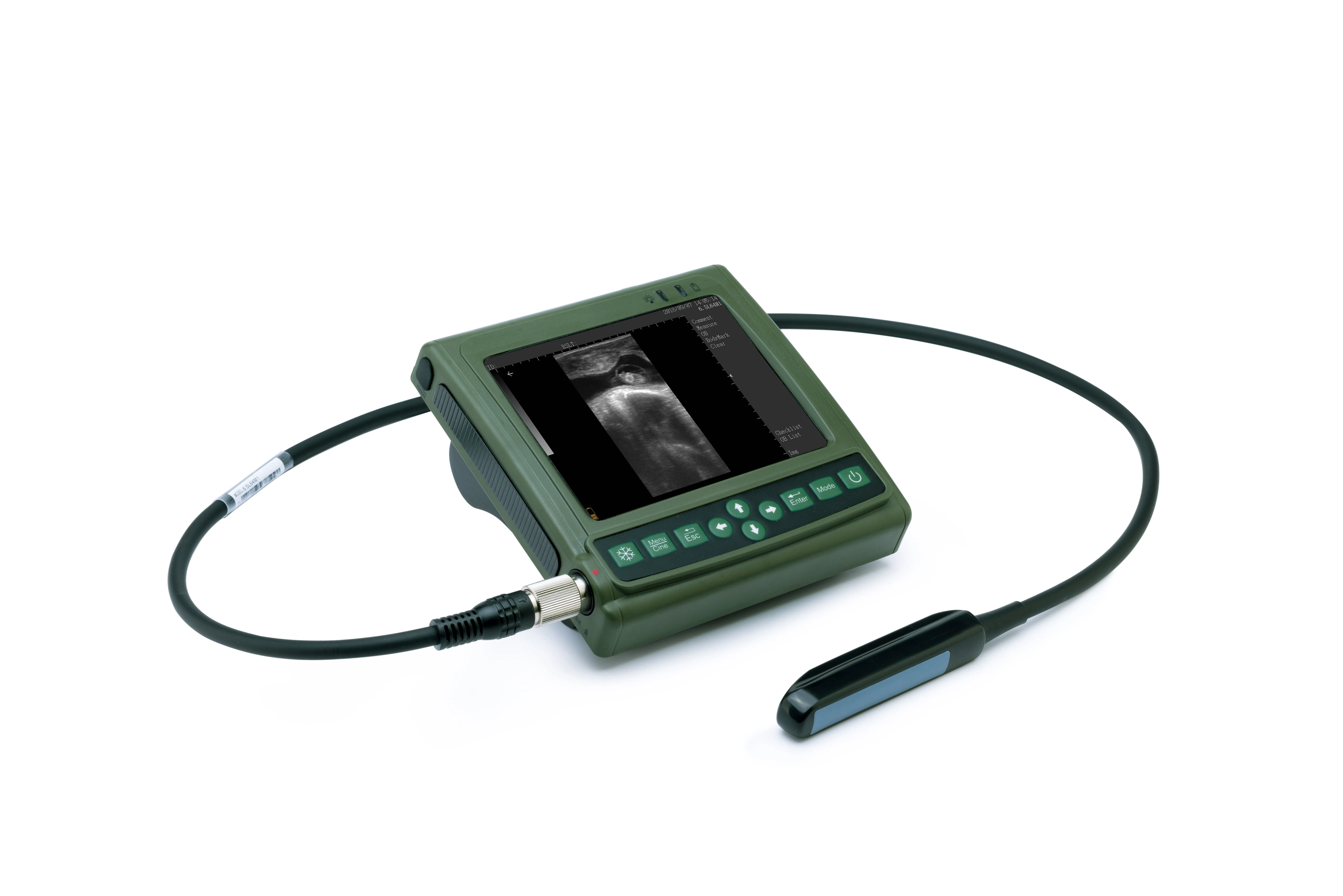
256 gray levels aren't just a number for marketing, they actually resolve the echogenicity difference between a renal cortex and medulla in a cat kidney at around 50mm depth (teh unit handles max 236mm across 10 grades of stepped adjustment), the B/M mode overlays a motion trace on the 2D image for cardiac cycles and runs at 30fps or so.
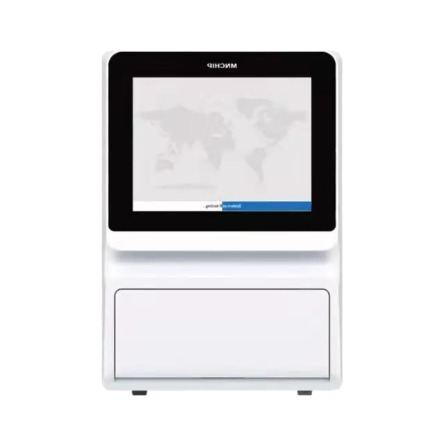
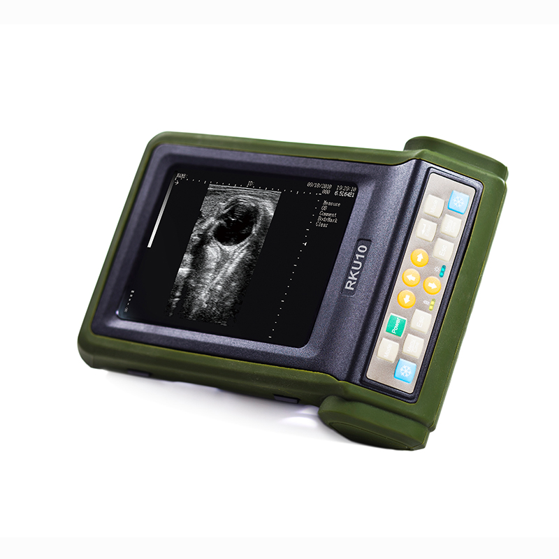
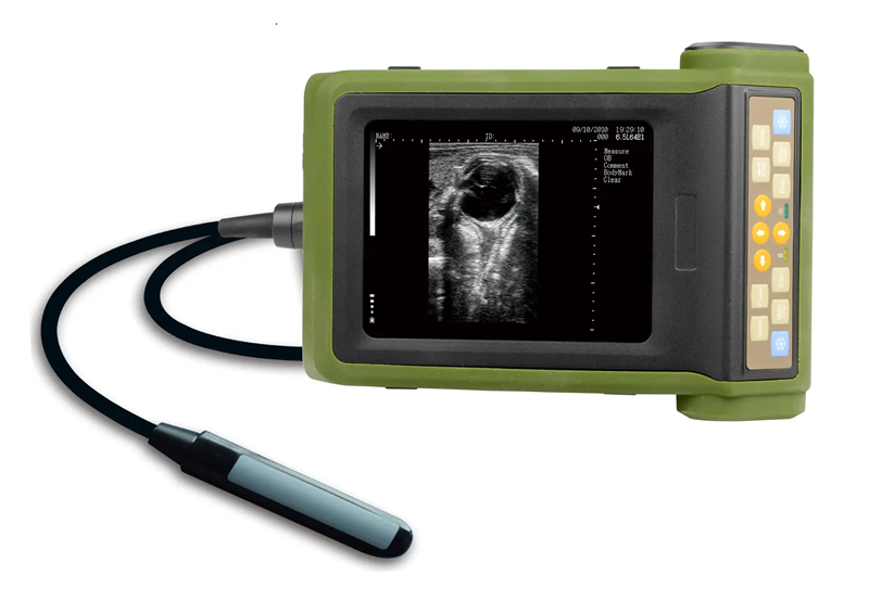
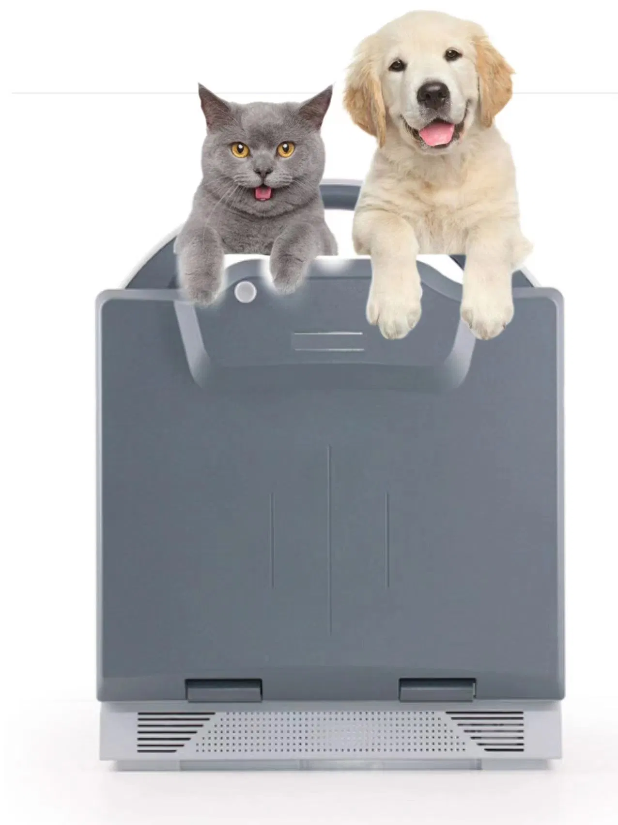
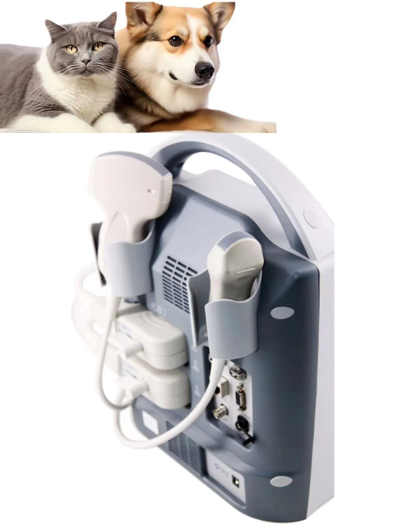
We've had it in the truck for six months now, the 10.4-inch LED color backlight holds up fine in direct sunlight but the 4GB onboard storage fills up fast—roughly 5000 frames—so you'll be offloading to USB every few days, also puncture guide bracket can be adjusted but the metal bracket is a bit finicky with 18G needles on the micro-convex probe.
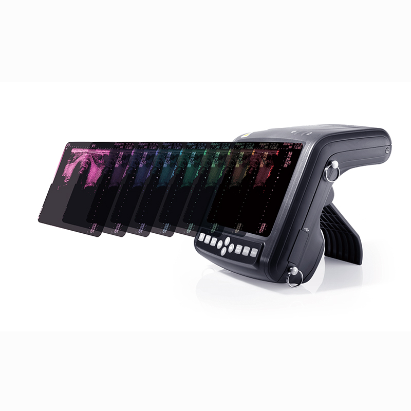
Actually the 8 pseudo-color maps are useful for differentiating fluid pockets in a splenic mass, just don't expect Doppler functionality because of that it only does B and M modes. 5. MOQ is usually 5 units for a custom laser engraving, otherwise single units ship form stock (check lead time on the 6.5MHz micro-convex replacements).
It gives you 256 levels of gray, which means smoother, more detailed tissue differentiation compared to lower-end systems. You’ll see finer contrast in soft tissues like liver or kidney, making diagnosis more reliable.
The TGC (Time Gain Compensation) has 8 adjustable steps that let you fine-tune the gain at different depths in real time. Each step is controlled via a slider on the interface, so you can quickly balance brightness from near to far field without digging through menus.
Yes, it has an adjustable puncture guide that works with the included 6.5MHz micro-convex probe. You can change the angle and position of the guide line on screen to match your needle approach, which is handy for biopsies or fluid drainage.
The internal 4GB holds about 5000 frames, and you can export images via USB or DICOM if your setup supports it. That way you’re not stuck with just the onboard storage—you can archive or share scans with colleagues.
They cover common species like dogs, cats, horses, cattle, and small exotics—each mark has labeled anatomical diagrams. There’s no option to add custom marks in the standard software, but the preset 27 cover most routine exams.
Great experience overall. The delivery was incredibly fast and efficient, arriving well ahead of the estimated schedule. Packaging was secure and professional, ensuring the product was perfectly protected during transit. The quality of the scanner itself appears to be excellent upon initial inspection; the build feels robust and the materials seem high-grade. Customer service was responsive and helpful during the ordering process, answering all my preliminary questions promptly. While I haven't had the chance to use it extensively in the field yet, the initial setup was straightforward. The logistical handling was seamless from warehouse to doorstep. I am very satisfied with the service support provided so far, as they were courteous and knowledgeable. The product's physical presentation matches the description perfectly. This transaction was handled with great care from start to finish. Genuinely impressed by the attention to detail in both the shipping process and the product's apparent craftsmanship. Good communication was maintained throughout, with timely updates on the shipment's progress. The overall service level has been commendable, making the purchase process worry-free. Getting the item so quickly was a pleasant surprise. The scanner arrived in pristine condition, which speaks volumes about their packaging standards. I have no complaints regarding the delivery or the initial quality assessment. The support team was proactive in ensuring a smooth experience. Great job on fulfilling the order accurately and swiftly. The logistics partner performed admirably, with clear tracking information provided. Overall, a very positive first impression based on the non-operational aspects of this purchase.
Outstanding experience from start to finish. The delivery was incredibly fast and the packaging was secure, ensuring the scanner arrived in perfect condition. The build quality feels robust and professional, exactly as described. Customer service was responsive and helpful when I had a pre-delivery question. A very smooth and satisfactory transaction overall.
Prompt delivery was impressive; the package arrived ahead of schedule and in perfect condition. Packaging was secure and professional, ensuring the device was well-protected during transit. Product quality appears excellent upon initial inspection, with a solid build and clear display. Customer service was responsive and helpful during the ordering process, answering all inquiries quickly. Overall, a very satisfactory experience from start to finish.
Keenly impressed with the prompt delivery; the logistics were handled seamlessly from start to finish. The packaging was exceptionally secure, ensuring the scanner arrived in pristine condition without any delays. Regarding quality, the construction feels robust and professional, exactly as described for veterinary use. The service provided was outstanding; the support team was responsive and helpful during the setup process. Overall, a very satisfactory experience that I would highly recommend to other professionals in animal care.
The delivery was exceptionally fast, arriving well ahead of the estimated schedule. Tracking information was precise and updated regularly, which made the entire process transparent and stress-free. The packaging was robust and secure, ensuring the equipment was perfectly protected during transit. There were no signs of damage or mishandling, reflecting a high standard of logistical care. Regarding the item itself, the build quality is immediately impressive, with a solid, professional feel that inspires confidence. The materials used seem durable and suited for clinical environments. Initial setup was straightforward, with clear documentation provided. As for customer support, the communication was prompt and helpful from the outset. Any pre-delivery inquiries were answered comprehensively and courteously. This level of service, combined with the efficient logistics and apparent product quality, makes for a very positive purchasing experience overall. The attention to detail in every step, from ordering to unboxing, is commendable and sets a high benchmark. It is clear that the seller prioritizes customer satisfaction and reliable service, which is crucial for professional equipment of this nature. The seamless integration of logistics, quality assurance, and responsive support is noteworthy. This holistic approach to the sale and delivery of a technical product is highly appreciated and certainly influences future purchasing decisions. The overall impression is one of professionalism and reliability, which are essential factors when acquiring diagnostic tools for veterinary practice. The transaction was smooth from beginning to end, with no complications or delays encountered. Such a dependable and efficient service model is invaluable, saving time and providing peace of mind. The seller’s commitment to excellence in both product and service delivery is evident and greatly valued.
Beyond expectations in every aspect! The delivery was incredibly fast, arriving well ahead of the estimated date, which was a pleasant surprise. Built with robust materials, it feels durable and reliable for professional use. Customer service was outstanding—responsive and helpful throughout the process. A truly seamless experience from order to receipt.
Yay, the delivery was incredibly fast and efficient, arriving well before the estimated date, which was a pleasant surprise. The packaging was secure and intact, ensuring the item reached me in perfect condition without any damage. Regarding quality, the build feels robust and durable, suggesting it can withstand regular use in a veterinary setting. The service from the seller was outstanding; they were responsive and helpful, promptly addressing my queries with clear and friendly communication. Overall, I'm thoroughly impressed with the seamless experience from order to receipt, and I'd highly recommend this for anyone in need of reliable veterinary equipment.
Logistics were impressively fast and seamless, with the package arriving ahead of schedule and in perfect condition. The quality of the scanner is outstanding, featuring durable construction and precise imaging that meets professional standards. Service was exceptional, with responsive and knowledgeable support that made the entire process smooth and hassle-free.
Delivery was incredibly fast and efficient, with the package arriving well-protected and ahead of schedule. Dependable shipping made the entire process smooth and hassle-free. Despite some initial concerns, the item's build quality is robust and meets professional standards, showing no defects upon inspection. The customer support team was responsive and helpful, promptly addressing my inquiries with clear and friendly communication. Definitely a reliable purchase overall, and I'm satisfied with the seamless experience from order to delivery.