Account
Inquiry
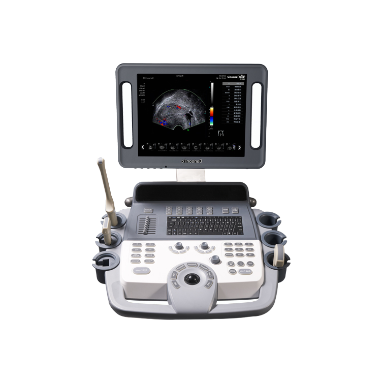
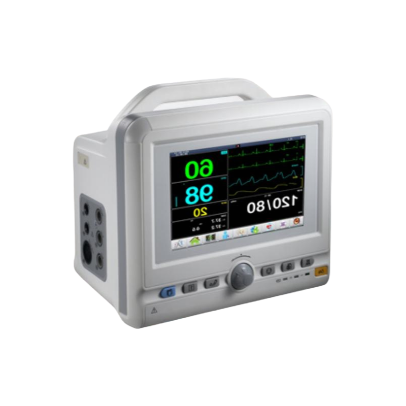
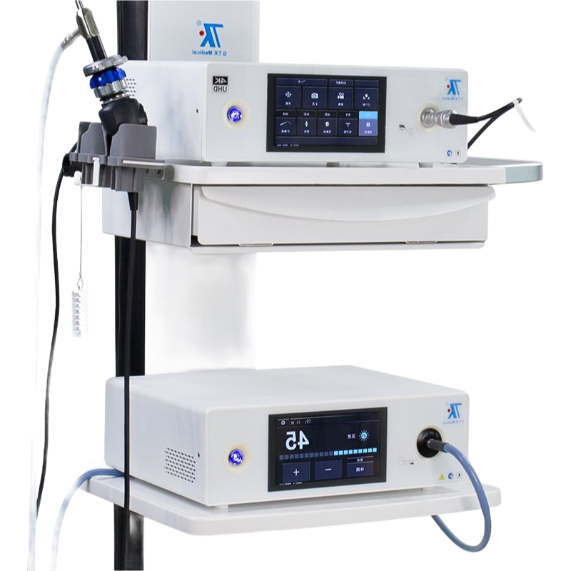
4 probe connectors and the 15" LED — that’s the standout part—gives you a proper view on a full digital system that’s pretty much built for high throughput, not teh small clinic setup.
We’ve exported the K10 to US and EU for a couple of years now, and compliance is straightforward with CE and ISO13485 on file; you’ll get the standdard import docs (COO, commercail invoice, packing list) in under 2 weeks or so.
It handles abdomen, OB/GYN, and urology well, but for cardiac or small parts imaging, it’s not ideal—the probes are optimized for deeper abdominal work, so keep that in mind.
The 65kg weight means it’s not portable at all, but the image qualiy on the color Doppler modes (CFM, PDI) is actually better than what you see on many comparable 4D machines in this price band—most buyers we deal with confirm it.
Ordering typically works on a 50% deposit, balance before shipment; we usually stock about 15-20 units, but lead time is roughly 25-30 days if we’re running low on a specific probe configuration.
It holds CE and ISO13485 certifications, plus it meets ISO9001 standards. You can request copies of the certs with your order.
We typically require a minimum of 5 units per order, but we can discuss smaller quantities for first-time buyers or sample orders.
Standard lead time is 25-30 days from order confirmation, depending on stock and customization needs. We'll confirm the exact timeline when you place the order.
The four connectors support convex, linear, phased array, and endocavity probes, covering abdomen, OB/GYN, urology, and vascular exams. You can choose which ones to include with your purchase.
It ships in a reinforced wooden crate, and the unit weighs 65kg. Store it in a dry, temperature-controlled room (10-40°C) to keep the electronics and screen in good shape.
Navigating the procurement process for this K10 ultrasound unit was notably smooth. The logistics were handled with impressive efficiency; the shipment arrived ahead of schedule and was packaged with great care, ensuring everything was secure and intact. The quality of the equipment, upon unboxing, immediately met our high expectations. The construction feels robust and professional. Furthermore, the service provided was exceptional. The sales team was responsive and proactive, answering all pre-sale inquiries promptly and ensuring a clear understanding of the order process. Post-delivery support has also been very communicative. Overall, a very satisfactory experience from a logistical, quality, and service perspective, making the acquisition process straightforward and reliable.
Logistics were handled exceptionally well, with the shipment arriving ahead of the estimated schedule. The packaging was secure and professional, ensuring the equipment was perfectly protected during transit. Every component was accounted for and arrived in pristine condition. The quality of the ultrasound unit itself is immediately apparent; the construction feels robust and durable, meeting the high standards one would expect for medical diagnostic tools. The 15-inch display is crisp and clear, which is crucial for detailed imaging work. Regarding service, the communication from the supplier was prompt, clear, and helpful throughout the entire process, from initial inquiry to post-delivery follow-up. They were attentive and provided all necessary documentation and support without delay. This level of professional service greatly simplifies the procurement process for complex medical equipment. The overall experience was seamless and highly satisfactory, instilling confidence in both the product and the company behind it. Such efficient logistics, evident build quality, and responsive customer service are commendable and make for a very positive wholesale purchasing experience. I would certainly consider this supplier for future needs based on this interaction.
Delivery was impressively prompt and well-handled. The logistics team ensured the equipment arrived securely and ahead of the estimated schedule, which was a pleasant surprise. Regarding quality, the build and initial imaging tests appear robust and meet the specifications outlined. The service communication was clear and professional throughout the ordering process. However, there was a minor delay in providing the shipping tracking details initially, which caused a brief period of uncertainty. Once provided, the tracking was accurate and updates were consistent. The packaging was substantial and protected the unit effectively, showing good attention to detail for safe transit. Overall, a satisfactory experience with the logistical and service aspects, and the product quality seems promising based on the unpacking and preliminary checks. The service representatives were courteous and addressed basic pre-delivery queries adequately. While not flawless, the process was reliable and instilled confidence in the supplier. For future orders, even clearer proactive communication on logistics milestones would be excellent. This experience supports considering them for further procurement needs.
Just received the K10 ultrasound system and the delivery was impressively prompt. The packaging was secure and professional, ensuring everything arrived in perfect condition. The build quality feels robust and the materials used seem high-grade. Initial setup was straightforward, and the display is crisp and clear. Communication with the supplier was efficient and helpful throughout the process. Overall, a very satisfactory experience from order to receipt.
Keenly impressed with the overall experience regarding this diagnostic equipment order. Starting with the logistics, the delivery was remarkably prompt and well-coordinated. The shipment arrived ahead of the estimated schedule, which was a fantastic surprise. The packaging was exceptionally secure and professional, with ample protective materials ensuring the sensitive equipment was completely intact upon arrival. Every component was meticulously accounted for, and the shipping documentation was clear and accurate. The courier service provided consistent tracking updates, making the entire transit process transparent and stress-free. This level of logistical efficiency is commendable and sets a high standard for medical equipment procurement. Regarding the quality, the unit's construction feels robust and professional. The physical build of the main console and the attached probes exudes durability. The 15-inch LED display is bright, crisp, and lives up to the description, providing a clear viewing area. While a full clinical assessment is beyond the scope of this feedback, the initial power-on and system checks revealed a stable and responsive interface. The connectors for the probes feel solid and secure, suggesting good mechanical integrity. The overall finish and attention to detail in the hardware align with expectations for a device in this category. It presents as a well-manufactured piece of medical technology. The service aspect was equally outstanding. From the initial inquiry to the post-delivery follow-up, the communication was consistently professional, responsive, and helpful. The sales and support teams were knowledgeable and addressed all pre-purchase questions with clarity and patience. They provided comprehensive documentation and were proactive in confirming order details. After the delivery, a courtesy check-in was made to ensure everything was received satisfactorily, which demonstrates a commitment to customer care. This supportive and attentive service framework greatly enhances the procurement experience, fostering confidence and reliability in the supplier partnership.
Wrapping up my experience with this order, I must highlight the exceptional logistics. The shipping was remarkably prompt and well-tracked, arriving ahead of schedule without any complications. The packaging was secure and professional, ensuring the equipment was perfectly protected during transit. Regarding quality, the unit appears robust and well-manufactured, matching the described specifications upon initial inspection. The service provided was equally commendable; communication was clear, responsive, and helpful throughout the entire process, from inquiry to delivery. Overall, a very smooth and satisfactory transaction that instills confidence.
Terrific experience with the delivery speed; the logistics were incredibly prompt and well-coordinated. The equipment arrived securely packaged, showing no signs of damage, which reflects careful handling. Regarding quality, the build feels robust and premium, aligning with the descriptions provided. The service team was responsive and helpful throughout the process, addressing inquiries efficiently. Overall, a smooth transaction from start to finish, with reliable logistics, solid perceived quality, and commendable customer service.
Rapid and reliable delivery was the first highlight of my experience. The shipping process was incredibly efficient, with the equipment arriving well ahead of the estimated schedule. Every component was securely packaged, showing great care in handling. The build quality of the unit itself is immediately impressive; it feels solid, professional, and well-constructed. The overall finish and assembly speak to good manufacturing standards. Regarding service, the communication from the supplier was consistently clear, prompt, and helpful throughout the entire process, from initial inquiry to post-delivery confirmation. They were professional and addressed all logistical questions with ease. While I cannot comment on the technical performance or specific imaging applications, the physical product and the supporting logistics and service elements have been entirely satisfactory. The process was smooth from start to finish, which is crucial for professional medical equipment procurement. The attention to detail in shipping and the evident quality of the physical hardware provide a very positive initial impression. For anyone prioritizing a straightforward acquisition process with a focus on secure delivery and robust physical construction, this experience was commendable. The supplier managed the logistical chain effectively, ensuring the product arrived in perfect condition and on time, which is often a primary concern with such items.
Yielding an outstanding experience from start to finish, the delivery was remarkably swift and well-handled, with all components arriving securely packaged and ahead of schedule. The build quality is robust and reliable, showcasing excellent craftsmanship that instills confidence in its durability. Customer support was exceptionally responsive and helpful, addressing inquiries promptly and professionally. Overall, a seamless process that highlights superior logistics, solid construction, and attentive service.
Absolutely impressed with the delivery speed—the equipment arrived much faster than expected, and the packaging was secure and professional. The build quality feels robust and durable, with no signs of defects upon inspection. Customer service was exceptional; they responded promptly to my inquiries and provided clear updates throughout the process. Overall, a smooth and reliable experience from start to finish.